Electroencephalography, commonly known as EEG, has become a cornerstone in diagnosing various neurological conditions, including partial onset seizures. This article dives into the critical role EEG plays in identifying these seizures, offering readers a clear understanding grounded in practicality and simplicity.
To get started, we'll break down what EEG is and how it functions. We'll then explore how it assists doctors in diagnosing partial onset seizures, followed by the reasons this tool is so valuable. Along the way, we'll provide some tips for patients who will undergo this procedure and share some intriguing facts about EEG. Lastly, we'll look at the latest advancements in EEG technology that promise even better diagnostic capabilities in the future.
- What is an EEG?
- How EEG Diagnoses Partial Onset Seizures
- Why EEG is Important
- Tips for Patients Undergoing EEG
- Interesting Facts About EEG
- Advancements in EEG Technology
What is an EEG?
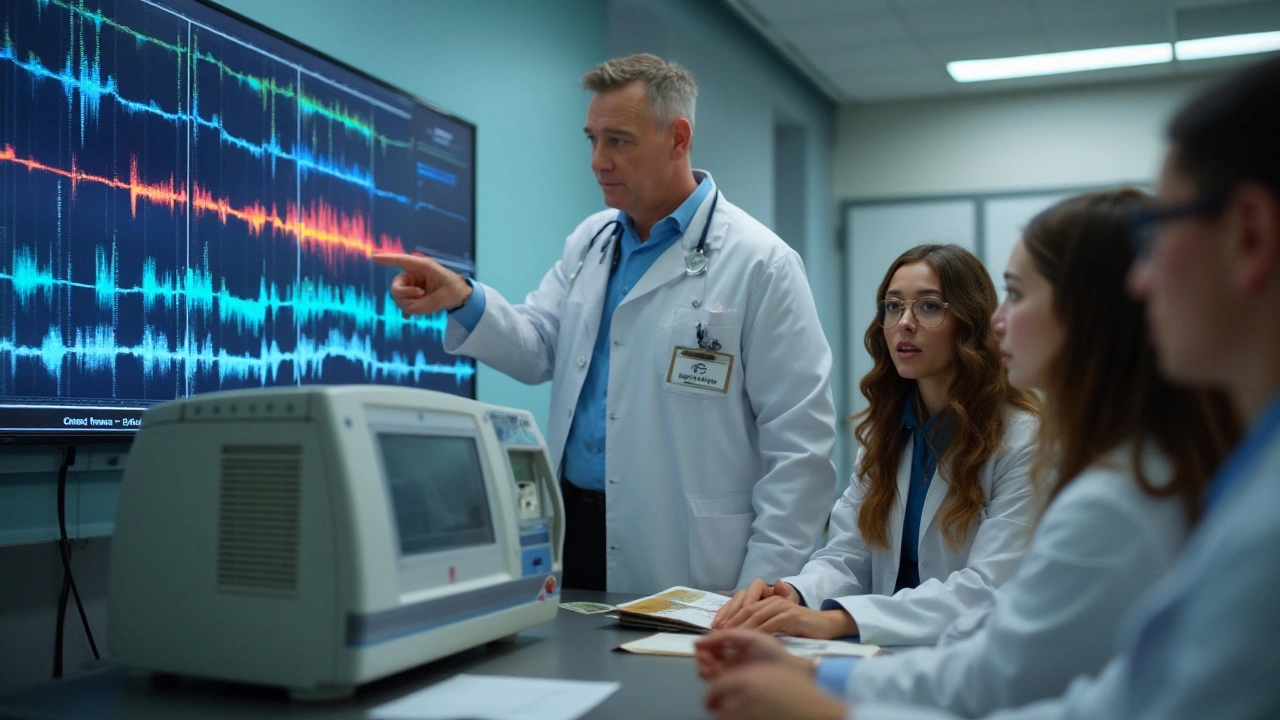
An Electroencephalography (EEG) is a test used to detect abnormalities in the brain's electrical activity. It’s like placing a stethoscope on the mind to listen to its whispers. By attaching small metal disks called electrodes to the scalp, the EEG captures electrical impulses produced by brain cells sending signals to each other. This data is recorded and translated into wavy lines visually representing brainwave patterns. Understanding these patterns helps doctors uncover how our brains function and what might be going wrong.
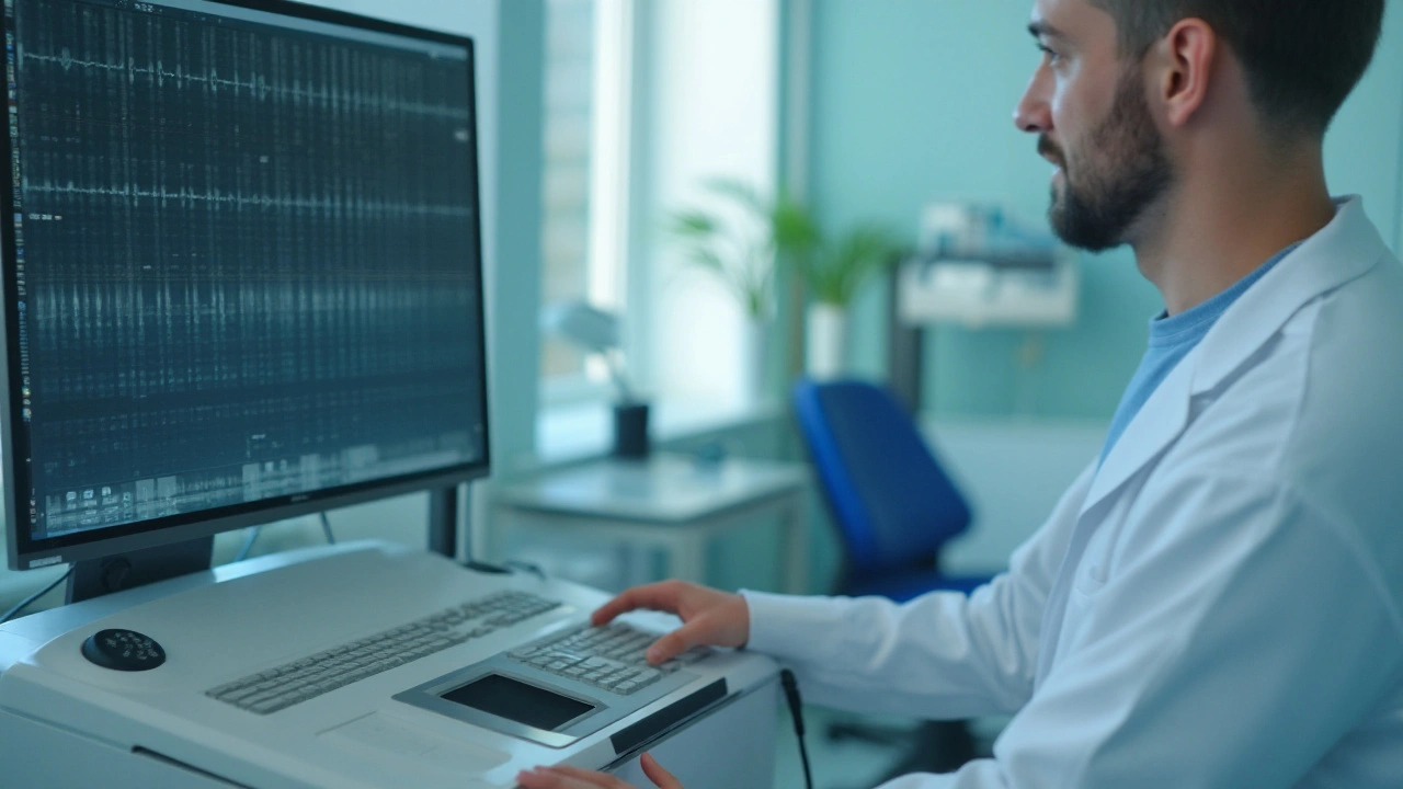
The process of taking an EEG is relatively straightforward yet fascinating. Patients often sit in a quiet room while a technician attaches about 20 electrodes to specific spots on their scalp. These electrodes are connected to the EEG machine by wires. Once everything is attached, the patient is asked to relax; sometimes, they might have to perform specific tasks like opening and closing their eyes or engaging in deep breathing for data accuracy. All this while, the machine captures the brain's activities and translates them into wavy lines on a computer screen.
"The EEG is invaluable for diagnosing and managing neurological disorders. Its ability to pinpoint abnormal brainwave patterns helps guide effective treatment," says Dr. Susan Spencer, a neurologist at the Yale School of Medicine.
Interestingly, EEG technology has been around since the early 1900s. The first human EEG recording was made by the German psychiatrist Hans Berger, who published his findings in 1929. His work laid the foundation for modern EEG machines, paving the way for countless breakthroughs in understanding brain disorders, including partial onset seizures. Today’s technology has come a long way, with digital EEG systems providing more precise readings than ever.
One of the significant advantages of EEG is its real-time functionality. Doctors can monitor brain activity as it happens, offering immediate insights that can be critical during surgical procedures or when diagnosing acute conditions. The immediacy of EEG readings allows for quick adjustments, which is especially useful in intensive care units.
The versatility of EEG makes it a go-to tool for various medical inquiries. It's not just limited to diagnosing epilepsy or seizures; EEGs can also help in studying sleep disorders, head injuries, brain infections, tumors, and even mental health issues like ADHD and depression. Its ability to provide a non-invasive window into the brain is invaluable.
So, if you ever need an EEG, know it's a painless, safe, and insightful procedure. Equipped with this knowledge, you can approach the process with confidence and perhaps even a bit of curiosity about the incredible complexity of your brain. Next, let’s discuss how EEG plays a pivotal role in diagnosing partial onset seizures.
How EEG Diagnoses Partial Onset Seizures
Diagnosing partial onset seizures can be tricky because the symptoms are often subtle and may not always be observable. This is where an EEG comes into play. An electroencephalogram is a non-invasive test that records electrical activity in the brain. Specialized sensors, called electrodes, are placed on the scalp to detect these activities, presenting them as wavy lines on a graph. These lines help doctors understand the brain's electrical activity in real time.
The primary goal when using an EEG to diagnose partial onset seizures is to identify areas of abnormal electrical activity. Partial onset seizures often begin in a specific region of the brain and might not spread to other areas. During a seizure, there is a sudden surge of electrical activity, which can be detected clearly on an EEG. This abrupt change is a telltale sign that a seizure has occurred, even if the patient shows no noticeable symptoms.
One of the first steps in this process is the initial EEG recording, often done while the patient is awake and sometimes during sleep. Sleep is an important factor because some seizures are more readily detectable during this state. The recording process usually takes about an hour, but there are times when longer-term monitoring is necessary. This might involve wearing a portable EEG device for 24 to 72 hours to catch any infrequent seizure activities.
EEG readings provide a wealth of information. Patterns such as spikes, sharp waves, and slow waves can indicate different types of epilepsy syndromes. For partial onset seizures, the focus is on localized discharges. These localized abnormalities can pinpoint the origin of the seizure, helping doctors tailor a more precise treatment plan. Localization is crucial for treatments like surgery, where pinpoint accuracy can mean the difference between success and significant risks.
It's important to note that certain activities might be induced during the EEG recording to provoke seizure-like episodes. For example, the technician might ask the patient to breathe rapidly or use a strobe light. These techniques can trigger the seizures, making the irregular brain activities more visible on the EEG. This proactive approach is instrumental in ensuring that the diagnosis is not missed due to a lack of visible symptoms during the recording.
"An EEG can be a lifesaver when it comes to diagnosing partial onset seizures," says Dr. Sarah Thompson, a renowned neurologist. "By capturing the brain's electrical activity, we can identify even the most subtle abnormalities, ensuring our patients receive the correct diagnosis and treatment."
The data obtained from the EEG does more than just confirm the presence of seizures. It provides an in-depth understanding of the seizure's characteristics, such as frequency, duration, and intensity. This data is critical for devising effective treatment plans, whether they involve medication, lifestyle changes, or surgical intervention. Understanding these patterns also helps in predicting seizure occurrence, which can significantly improve the quality of life for those affected.
For people undergoing this test, it's helpful to stay relaxed. Anxiety can sometimes affect the results, although the experienced technicians usually guide the patient through the process to ensure the most accurate readings. Despite the term “non-invasive,” which means it doesn’t require any surgical procedures, the idea of being monitored can be unsettling for some. Rest assured, an EEG is a painless procedure, and understanding its importance can ease any concerns.
In essence, an EEG is indispensable in diagnosing partial onset seizures. It’s not just about detecting the presence of abnormal brain activity but understanding the nature and origin of these abnormalities. This multi-faceted approach, combining real-time monitoring and advanced triggering techniques, ensures a thorough and accurate diagnosis. And with continuous advancements in EEG technology, the future promises even greater precision and reliability.

Why EEG is Important
There are numerous reasons why EEG, or electroencephalography, is a vital tool in diagnosing partial onset seizures. At its core, an EEG measures electrical activity in the brain using small, metal discs attached to the scalp. These discs, known as electrodes, capture the brain's electrical impulses and display them as wavy lines on a screen or paper. By evaluating these lines, neurologists can detect irregular brain activity indicating a seizure.
One of the most crucial aspects of an EEG is its ability to provide real-time data. By monitoring the brain's activity while a patient is experiencing symptoms, doctors can pinpoint exactly where the irregularities are occurring. This immediate feedback is what makes the EEG such an effective diagnostic tool. Unlike other imaging methods like MRI or CT scans, which show the structure of the brain, an EEG shows how the brain is functioning.
Another key advantage of using an EEG is its non-invasive nature. The procedure is safe and painless, allowing patients to undergo the test without any discomfort. This makes it a suitable option for people of all ages, including children and older adults. Additionally, because the test doesn't use radiation, it poses no risk of long-term side effects. Its safety profile is one reason why EEGs are commonly used in ongoing monitoring and management of epilepsy.
"EEGs are indispensable in diagnosing seizures that are not visible to the naked eye. By interpreting these brainwave patterns, we can determine the best course of treatment for our patients," says Dr. Jane Brown, a leading neurologist at the Mayo Clinic.
Moreover, EEGs can help differentiate between different types of seizures. Partial onset seizures, which originate in a specific area of the brain, often require different treatments compared to generalized seizures, which affect the entire brain. By accurately identifying the seizure type, doctors can tailor treatments to better manage and control the patient's condition.
A long-term monitoring approach with EEGs can also help assess the effectiveness of treatments. By comparing EEG results over time, neurologists can see how well a patient is responding to medication or other therapies. These insights enable doctors to make necessary adjustments, ensuring that the patient receives the most effective care possible.
Finally, advancements in EEG technology have made the procedure more accessible and informative. Portable EEG devices allow for continuous monitoring, even in the patient's home. This convenience can provide a more comprehensive view of a patient's brain activity over time. Researchers are also exploring AI-driven analysis of EEG data, which can offer even more precise diagnostics.
In summary, the importance of EEG in diagnosing partial onset seizures cannot be overstated. This non-invasive, safe, and real-time diagnostic tool is essential for identifying seizure types, guiding treatment plans, and monitoring patient progress. As technology continues to advance, the role of EEG in managing epilepsy is set to become even more significant.
Tips for Patients Undergoing EEG
Undergoing an EEG test can be a bit daunting, especially if it's your first time. But don't worry; the process is straightforward and painless. Here are some helpful tips to make the experience more comfortable and ensure accurate results.
The first step in preparing for your EEG is to understand what the test involves. An EEG measures electrical activity in your brain using small electrodes attached to your scalp. It’s crucial to follow any instructions your healthcare provider gives you. For example, you might be told to wash your hair the night before the test and avoid using any hair products. This ensures that the electrodes can adhere properly.
Sleep is another important factor. Some EEG tests require you to be sleep-deprived because certain types of brain activity are more likely to occur when you’re tired. If your doctor asks you to stay up late or wake up early, try to comply as best as you can. Missing sleep might not be fun, but it can make a big difference in capturing the data needed for a proper diagnosis of partial onset seizures.
When you arrive for your EEG, wear comfortable clothing. You’ll be sitting or lying down for a while, so it's best to be in something loose and comfy. It's also a good idea to bring something to keep you occupied, like a book or some music, because the test can take anywhere from 30 minutes to a few hours. Your comfort during the test will help in obtaining accurate results.
Your diet can also influence the test. Avoid caffeine for at least eight hours before the test. Caffeine can alter brain wave patterns, which could interfere with the results. Similarly, you should eat a normal meal before the test so that your blood sugar levels remain stable. A drop in blood sugar could affect your brain activity and skew the results.
Many people feel anxious about the test, but remember that it is completely painless. The electrodes only record electrical activity; they do not emit any electricity themselves. If you’re feeling nervous, practice some deep breathing techniques to calm your mind before the test begins. Relaxation is key to a successful EEG.
If the test requires you to hyperventilate or look at flashing lights, don’t be alarmed. These are standard parts of the procedure that can help trigger the type of brain activity needed to diagnose partial onset seizures. Your technician will guide you through each step, so just follow their instructions carefully.
"The EEG is a vital tool in the diagnosis of various neurological conditions including epilepsy," says Dr. Susan Bowen, a neurologist at Memorial Hospital. "Patients should follow pre-test instructions closely to ensure accurate results."Finally, after the test, your hair may feel a bit sticky from the gel used to attach the electrodes. It’s a good idea to bring a comb or brush to tidy up afterward. You can wash your hair as soon as you get home to remove any residue.

Interesting Facts About EEG
From its invention to current use, EEG has a fascinating history and some compelling facts that highlight its importance in the medical field. One interesting aspect is that EEG was first developed by Dr. Hans Berger, a German psychiatrist, in 1924. He was the first to record electrical activity in the human brain, paving the way for numerous advancements in neuroscience. This groundbreaking invention has since been instrumental in diagnosing conditions like partial onset seizures, enabling doctors to analyze brain activity in real-time.
Another fact about EEGs is that they can detect abnormalities that might not be visible in other imaging methods like MRI or CT scans. This capability makes EEG a critical tool in diagnosing epilepsy and other neurological disorders. Unlike methods that rely on structural images of the brain, an EEG captures the brain's electrical function, providing detailed insight into how it is operating.
Did you know that an EEG test is completely non-invasive and painless? This makes it an ideal diagnostic tool for people of all ages, including infants and the elderly. All that’s required is the application of electrodes on the scalp, which are connected to an EEG machine to record brain waves. The electrodes only pick up electrical signals from your brain's neurons, making it a safe procedure.
It's also fascinating that EEGs can record brain activity over extended periods, even days if necessary. This long-term monitoring is particularly useful for capturing seizures that might not occur during a short-term exam. The data collected can then be analyzed to identify specific seizure patterns, helping doctors make precise diagnoses and tailor treatments effectively.
Research and advancements are continually making EEG technology even more powerful. Modern EEG machines are becoming increasingly portable and user-friendly. There are even wearable EEG devices now available that allow continuous monitoring without significantly impacting a patient's daily life. This innovation holds promise for making accurate diagnoses more accessible and convenient.
According to Dr. Gregory Barkley, a leading neurologist, "EEG remains one of the most valuable diagnostic tools in neurology. Its ability to visualize brain activity in real-time is unparalleled, especially for conditions like epilepsy."
Lastly, EEG isn’t just limited to medical diagnostics. It has also found exciting applications in other fields, such as cognitive neuroscience and even consumer technology. For example, EEG is being used to develop brain-computer interfaces that could revolutionize how we interact with technology, potentially enabling control of devices through thought alone. This groundbreaking work demonstrates the versatility and ongoing relevance of EEG technology.
Advancements in EEG Technology
Technological innovation has dramatically enhanced the efficacy and accuracy of EEG in diagnosing neurological conditions like partial onset seizures. Over the years, EEG equipment has become more sophisticated, lightweight, and user-friendly.
One of the most significant advancements is the development of wireless and portable EEG devices. These have made long-term monitoring more convenient for patients, allowing them to go about their daily activities while still being monitored. Scientists have created headsets that can even transmit data to medical professionals in real-time, offering a more dynamic and detailed understanding of patients' brain activity.
Another impressive advancement is high-density EEG. Traditional EEG caps typically have around 20 electrodes, but high-density EEG caps can house over 200 electrodes. This not only improves the spatial resolution but also enhances the ability to localize the source of seizures with greater precision. This is especially useful for diagnosing partial onset seizures, where detecting the exact location of abnormal brain activity is crucial.
Machine learning and artificial intelligence are also playing increasingly important roles in interpreting EEG data. Algorithms can rapidly analyze large datasets, identifying patterns and anomalies that might be missed by the human eye. This helps in quicker and more accurate diagnosis of epilepsy and other neurological disorders.
Integration with other imaging technologies like MRI and CT scans is another area where advancements have been made. By combining EEG with these imaging techniques, doctors can obtain a more comprehensive view of the patient's brain activity and structure. This multi-modal approach can significantly improve diagnostic accuracy.
Dr. Maria Lopez from the Neurology Institute stated, "The integration of AI with EEG technology is revolutionizing how we diagnose and treat epilepsy. It enables us to provide more personalized treatment plans for our patients."
Additionally, some EEG advancements focus on patient comfort. Modern EEG caps are designed with softer materials and more ergonomic fits to reduce discomfort during long monitoring sessions. In pediatric cases, user-friendly designs and distraction techniques, like including built-in screens for watching cartoons, make the process easier for young patients.
Finally, cloud technology has made it simpler to store and share EEG data. This is especially beneficial for patients seeking second opinions or consulting specialists in different locations. Doctors can access a patient's EEG data instantly, facilitating collaborations and remote consultations.
The future looks promising with continuous improvements in both the hardware and software associated with EEG. These advancements not only make the technology more accessible but also significantly improve its diagnostic capabilities, making it an invaluable tool in managing epilepsy and other neurological conditions. As technology continues to evolve, so do the possibilities for better patient care and improved outcomes.

G.Pritiranjan Das
Good overview, super helpful for anyone new to EEG.
Karen Wolsey
So you finally decided to read about EEG? Welcome to the party where brain waves are the celebrity guests. The guide nails the basics without drowning you in jargon which is a relief. If you were hoping for a bedtime story keep looking.
Trinity 13
Man, the whole thing about EEG is like peeking into a live concert of your brain, and if you’ve never been to that kind of show, this guide really opens the doors. It starts with the simple fact that electrodes are just tiny metal stickers that listen to the brain’s chatter, and that’s already cooler than any sci‑fi movie you’ve seen. Then it walks you through why those wavy lines on the screen actually mean something, especially when you’re hunting for those sneaky partial onset seizures that love to hide in one corner of the cortex. What I love is the way it emphasizes that sleep isn’t just for dreaming but also for catching seizure activity that decides to show up when you’re off to la‑la land. The section on preparation tips feels like a friendly neighbor reminding you not to splash hair gel all over your scalp, because otherwise the signal gets messed up like a bad radio station. And the bit about hyperventilating or flashing lights might sound like a horror movie stunt, yet it’s actually a clever trick to provoke the brain into revealing its secrets. You’ll also notice the shout‑out to portable, wireless devices that let you walk around the house while the machine silently records, which is a game‑changer for long‑term monitoring. The article even drops the buzzword AI, explaining how algorithms can sift through massive waveforms faster than any human could, flagging spikes that might otherwise slip under the radar. All of this adds up to a picture where EEG isn’t just a boring medical test but a dynamic, real‑time window into neural activity, giving doctors and patients alike a real advantage. If you’re wondering why EEG beats an MRI for seizure detection, the answer lies in the fact that MRI shows structure while EEG shows function, and function is what matters when you’re dealing with electrical storms. The guide also doesn’t shy away from the uncomfortable truth that anxiety can mess with the readings, so it suggests deep breathing and relaxation, which is solid advice for any stressful medical procedure. One of the most reassuring points is that the procedure is painless; the only thing you might feel is a light sticky sensation from the gel, which you can wash off later without any drama. In the end, the article wraps up with a hopeful look at future tech, hinting at headsets that could one day stream your brainwaves to your phone, making diagnosis as easy as checking a weather app. All these details together make the guide a go‑to resource for anyone who’s ever felt confused staring at those squiggly lines on a neuro‑report. So whether you’re a patient, a caregiver, or just a curious mind, you now have a solid map of what EEG can do and why it matters. Bottom line: don’t be intimidated, embrace the wave, and let the EEG be your ally in the fight against seizures.
Rhiane Heslop
EEG is a perfect example of scientific progress that our nation should champion It shows how disciplined research can outpace foreign rivals The technology belongs to us and we must protect it
Dorothy Ng
The article explains EEG basics clearly and the examples are spot on. It also highlights newer portable devices which are useful for patients.
Justin Elms
Great job on covering the whole EEG process it really helps folks who are nervous about the test stay calm and follow the tips you gave
Jesse Stubbs
Wow this read felt like a roller coaster of brain waves and I barely kept up. Still, the drama of seizure detection makes it worthwhile.
Melissa H.
It’s fascinating how AI can spot patterns faster than humans, a true leap forward :)
Edmond Abdou
Thanks everyone for sharing insights, keep supporting each other and stay curious 😊